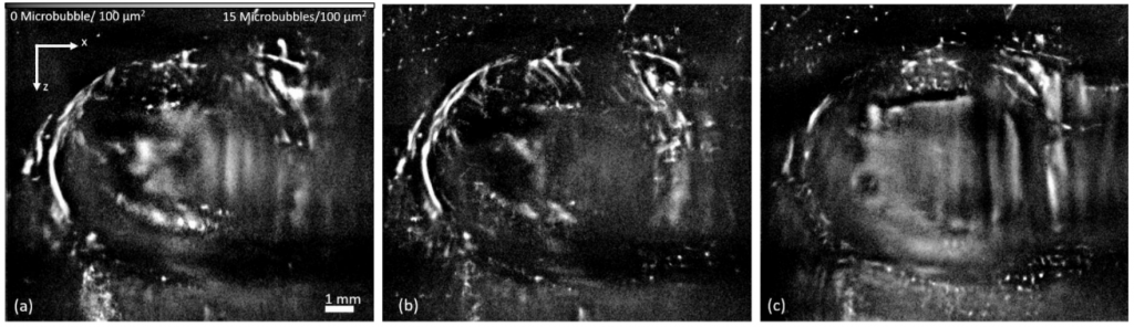
Ultrasound localization microscopy allows imaging the blood flow in organs in a fast, non-invasive and non-ionizing way. Based on the detection of microbubbles injected into the bloodstream, it is possible to obtain ultra-resolution angiograms of organs. However, conventional methods are based on the accumulation of microbubble positions detected during an acquisition period of about ten minutes. These methods do not allow the observation of moving organs. In order to map the vascular and microvascular network in the heart, it is therefore necessary to compensate for these movements which can have different origins: cardiac cycle, breathing, etc. For this purpose, we have developed MULA: an approach to correct the motion in the cardiac tissue during imaging and to accumulate the different positions of the detected microbubbles. This approach still needs to be extended in 3D to model the movements outside the imaging plane and obtain high resolution cardiac angiograms.
Our other projects
Cardiac angiography
Development of new dynamic biomarkers for the brain